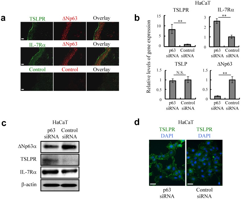
Figure 1. Expression of TSLPR and IL-7Rα in epidermal keratinocytes is controlled by ΔNp63.
(a) Immunofluorescence labeling confocal microscopy to localize TSLPR and IL-7Rα in the area of epidermis where ΔNp63 was not detected. Bar = 20 µm. (b, c) Quantitative RT-PCR (b) and immunoblot analysis (c) against ΔNp63, TSLPR, IL-7Rα and TSLP in control siRNA (siControl) and p63-specific siRNA (sip63)-transfected HaCaT keratinocytes. β-actin was used as a loading control. The cells were harvested for assays at 72 hr after siRNA transfection. Student's t test. **P<0.01 and N.S., not significant. (d) Immunofluorescence labeling fluorescence microscopy to localize TSLPR in siControl and sip63 HaCaT keratinocytes. The cells were fixed at 72 hr after siRNA introduction. DAPI, 4′,6-diamidino-2-phenylindole. Bar = 50 µm. Data are representative of at least three independent experiments.

