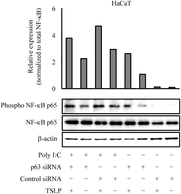
Figure 4. TSLP induces signal transduction in ΔNp63-deficient HaCaT keratinocytes.
Immunoblot analysis demonstrating phosphorylation of the p65 subunit of NF-κB at 24 hr after treatment with 10 µg/ml poly I:C and exogenous TSLP (10 ng/ml). The histogram shows the relative phospho- NF-κB expression normalized to NF-κB as determined by densitometric analysis. β-actin was used as a loading control. Data are representative of three independent experiments.

