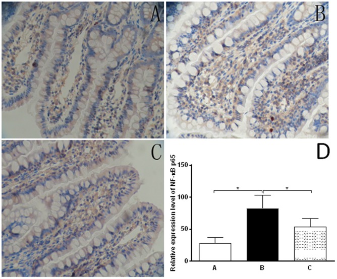
Figure 5. The immunohistochemical observation of stained NF-κB p65 in rat ileal tissues from different groups (SP×400).
The ileal sections were firstly de-paraffinized in xylene and rehydrated and antigen was retrieved by microwave and endogenous peroxidase activity was blocked by 3% H2O2 in methanol. The sections were probed with specific polyclonal rabbit anti-rat NF-κB p65 serum and the slides were then washed with PBS and incubated with respective secondary antibody and 0.1% diaminobenzidine substrate. Ten fields were randomly selected (400× magnification) and the results were quantitated. A: blank (normal) group; B: CCl4-treated rats receiving the vehicle only group; C: oxymatrine treatment group; and D: Bar graph showing the relative expression level of NF-κB p65 in rat ileal tissues. *P<0.01 by one-way ANOVA.

