
Figure 2.
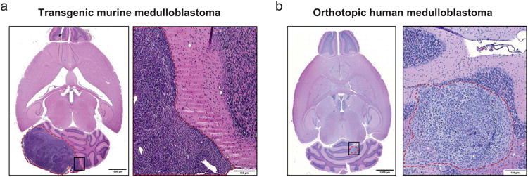
Experimental mouse models of medulloblastoma. (a) Transgenic mouse medulloblastoma 6 weeks after intracerebellar injection of retroviruses carrying Shh and Mycn (H & E). Right panel is enlarged image of boxed area in left panel. (b) Orthotopic human medulloblastoma (Daoy) xenograft 5 weeks after intracerebellar injection into nu/nu mice (H & E). Right panel is enlarged image of boxed area in left panel. Dashed red lines outline the tumor. Scale bars: 1500 μm (a, b left panels), 150 μm (a, b right panels).