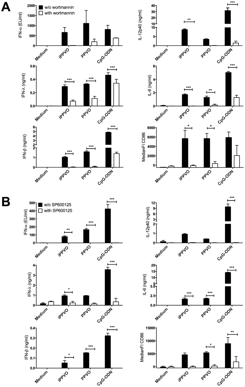
Figure 4. Inhibition of PI3K and JNK signalling greatly diminishes activation of pDC by PPVO.
BMDC of WT mice were generated and purified as described in material and methods. Purified pDC were stimulated in triplicates with the indicated stimuli in the absence or presence of PI3K inhibitor wortmannin (A) or JNK inhibitor SP600125 (10 µM) (B) and supernatants and cells were harvested after 24 h. Cytokine concentrations were determined in culture supernatants using ELISA and are depicted as mean +/− SEM. Surface expression of CD86 was analysed by flow cytometry and is depicted as mean +/− SEM of the median fluorescence intensity. Cytokine concentrations and CD86 expression were statistically analysed using 2-way ANOVA and Bonferroni post-test and differences are indicated by asterisks. One representative experiment of at least three is presented for cytokine data. Pooled data of two (A) or three (B) experiments are shown for CD86 expression.

