
Figure 2. Electrocardiogram (ECG) lead II recordings from NTG, Gαq-TG, and Gαq-TG+olmesartan mice.
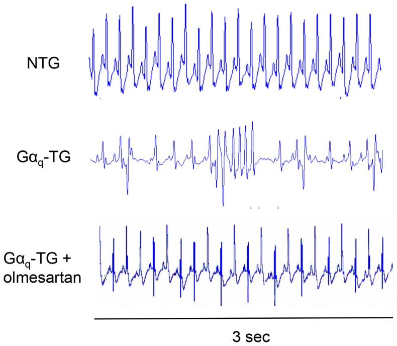
The middle ECG shows ventricular arrhythmias recorded from vehicle-treated Gαq-TG mice. PVC was frequently observed. In contrast, the upper and lower ECGs recorded from an NTG and olmesartan-treated Gαq-TG mouse showed P waves and QRS complexes with regular RR intervals without any arrhythmia, indicating a sinus rhythm. Mice at the age of 32 weeks were used.