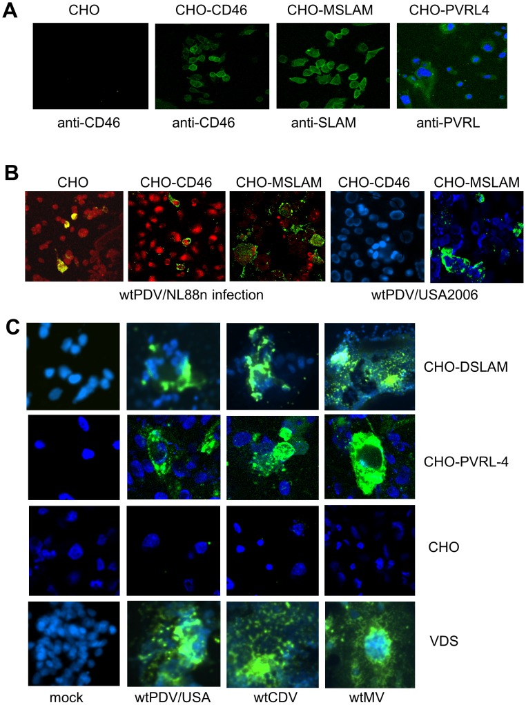
Figure 4. Increased infection of wtPDV infection in CHO cells expressing either SLAM or PVRL4.
(A) CHO-CD46, CHO-MSLAM and CHO-PVRL4 cells were stained with their respective receptor antibodies or mouse isotype control, fixed and stained with rabbit anti-mouse FITC. CHO cells were stained in the same manner with anti-CD46 antibody (B) CHO, CHO-CD46, and CHO-MSLAM cells were infected with wtPDV/NL88n and wtCDVUSA2006 (C) CHO, CHO-DSLAM, CHO-PVRL4 and VDS cells with wtPDV/USA2006, wtCDV and wtMV at MOI of 0.1 for 2 days. Cells were, permeabilised, fixed and stained with SSPE serum followed by rabbit anti-human FITC. Nuclei were either stained with propidium iodide and images take in a Leica TCS/NT confocal microscope or stained with DAPI and images taken using a Nikon Eclipse TE2000-U UV microscope (x400).

