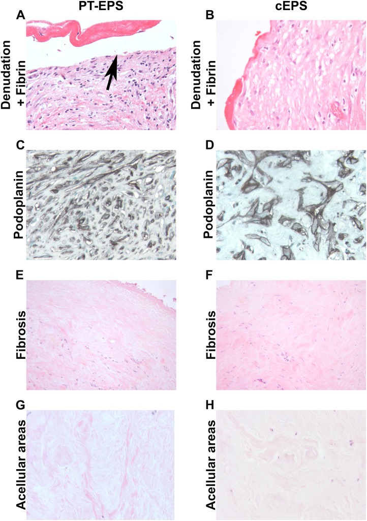
Figure 2. Morphologogical evaluation of peritoneal biopsies in PT-EPS and cEPS.
Peritoneal biopsies were either stained with PAS (A, B, E–H) or by immunohistochemistry with a monoclonal antibody against podoplanin (D2-40, C, D, original magnifications X 400 in A–D, G, H, X200 in E, F). The morphological evaluation demonstrated similar degrees of denodation and fibrin deposition (A, B), podoplanin positive cells (C, D), fibrosis (E, F) and acellular ares (G, H). (left column post transplant EPS; PT-EPS, right column classical EPS, cEPS).

