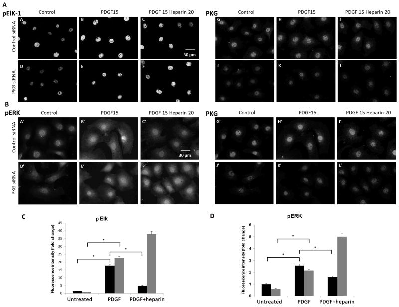
Figure 7.
Knockdown of PKG decreases heparin effects on ERK activity and pElk. A7r5 cells were electroporated with siRNA designed to knock down PKG in rat cells or scrambled RNA, and the cells were allowed to proliferate in growth media for 72 h with feeding at 24 h. At 72 h, cells were untreated, treated with PDGF for 15 min or heparin for 20 min before PDGF was added for 15 min. Panel A illustrates the pElk levels (pictures A–F). Staining for PKG is illustrated in the same cells in pictures G–L. Scrambled siRNA (A,B,C,G,H,I) as compared to PKG siRNA (D,E,F,J,K,L) is shown for cells not stimulated (A,D,G,J), PDGF treated cells (B,E,H,K) and heparin plus PDGF (C,F,I,L). Panel B illustrates an experiment where pERK was monitored (A′–F′) PKG staining for these cell samples is shown in pictures G′–L′. The treatment pattern is identical to that for panel A. These experiments are representative of two similar experiments each. Panel C provides quantitative analysis of Elk activation in PKG and scrambled siRNA. More than 100 cells from two separate experiments were analyzed and fluorescent intensity of pElk staining in the nucleus was determined. In both control (black bars) and PKG (grey) siRNA, pElk staining was significantly increased (p < 0.05,*). Heparin significantly (p<0.05,*) decreased pElk staining in control (black), but actually further increased pElk in PKG siRNA-treated cells. Panel D illustrates ERK activation as described for Elk in panel C. Nuclear pERK was determined for more than 100 cells from two experiments. As with Elk phosphorylation, PDGF increased ERK phosphorylation significantly (p<0.05,*) for both control siRNA (black bars) and PKG siRNA (grey bars) treated cells. In addition, heparin decreased PDGF-induced activation of ERK significantly (p<0.05,*) in control siRNA-treated cells, but not in PKG siRNA-treated cells.

