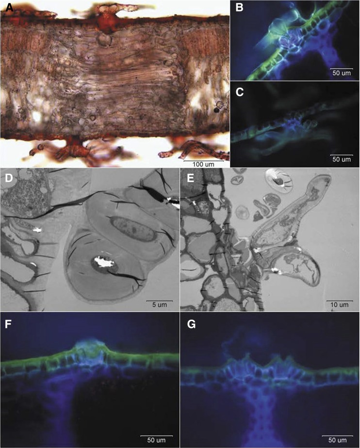
Figure 3.
Optical and TEM micrographs of intact adaxial and abaxial surfaces of holm oak young (A–F) and mature (G) leaves. A, Transversal section of a young leaf stained with Sudan IV. B, Adaxial leaf trichome. C, Abaxial leaf trichome. D, Base of an adaxial leaf trichome observed by TEM. E, Base of an abaxial leaf trichome observed by TEM. F, Detail of scar on a young, adaxial leaf surface after trichome shedding. G, Detail of a scar on a mature, adaxial leaf surface after trichome shedding. Note that the bases are near the bundle sheath extension (dark blue).

