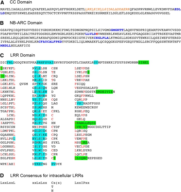
Figure 2.
Structural domains of the Rpg1r protein. A, The CC domain. The predicted CC is printed in orange, with the hydrophobic residues at the a and d positions in the heptad repeats underlined. The conserved EDVID motif (Rairdan et al., 2008) is printed in blue. B, The NB domain. Shown is the region corresponding to the known structure of the human Apaf1 NB-ARC1-ARC2 region. Previously described conserved motifs (van der Biezen and Jones, 1998; Meyers et al., 1999) are printed in blue in the following order: P-loop (kinase 1a), kinase 2, GLPL, RNBS-D, and MHD. An unusual substitution with a nonacidic residue within the kinase 2 motif is printed in red. C, LRR domain. Regions predicted to form α helices and β strands are highlighted in green and blue, respectively. Residues matching the consensus for intracellular LRRs are printed in red. D, Consensus for intracellular LRRs (Jones and Jones, 1997), where x can be any residue and L can be substituted with I, VM, or F.

