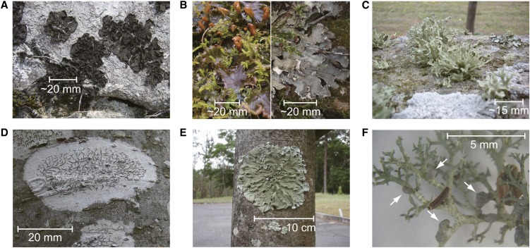
Figure 1.
Lichens analyzed in this study. A, Cyanolichen C. subflaccidum on a rock. B, Wet (left) and dry (right) thalli of cyanolichen Peltigera degenii with green moss. C, Chlorolichen R. yasudae on a rock. D, Chlorolichen Graphis spp. on a Zelkova serrata tree trunk. The grayish basal part of Graphis spp. is the site where the photobiont resides, and the dark-colored streaks are the apothecia. E, Chlorolichen Parmotrema tinctorum on a Z. serrata tree trunk. F, Cephalodia-possessing lichen S. sorediiferum. Some cephalodia are indicated by arrows. The stem- and branch-like structures are the green algae-containing compartments.

