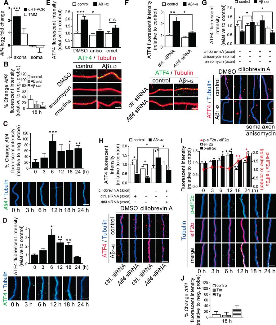
Figure 3. Atf4mRNA is recruited into Aβ1–42-treated axons, and axonal ATF4 protein is locally synthesized and retrogradely transported.
(A) Log2 fold change for Atf4 mRNA as determined by real time RT-PCR and DESeq2 (TMM). ***p<0.001.
(B) Hippocampal neurons were cultured in microfluidic chamber for 9–10 DIV, axons were treated with vehicle, Aβscrambled or Aβ1–40 for 18 h, and axonal Atf4 mRNA levels were measured by quantitative FISH. Mean ±SEM of 25–30 optical fields per condition (n=5–6 biological replicates).
(C) Axons were treated with Aβ1–42 for the indicated times, and axonal Atf4 mRNA levels were measured by quantitative FISH. Mean ±SEM of 25–40 axonal fields per condition (n=5–8 biological replicates per group). The background fluorescence was determined using a non-targeting probe (neg. probe) and set to zero. *p<0.05; **p<0.01; ***p<0.001. Scale bar, 5 µm.
(D) Neurons were cultures and treated as in C. Axonal ATF4 protein levels were measured by quantitative immunofluorescence. Mean ±SEM of 20–40 axonal fields per condition (n=4–8 biological replicates per group). *p<0.05; **p<0.01. Scale bar, 5 µm.
(E) Hippocampal neurons were cultured and treated as in B. 3 h prior to sample processing axons were treated with DMSO, anisomycin or emetine. Axonal ATF4 protein levels were determined by quantitative immunofluorescence. Mean ±SEM of 25–35 axonal fields per condition (n=5–7 biological replicates per group). ***p<0.001: *p<0.05. Scale bar, 5 µm.
(F) Hippocampal neurons were cultured in microfluidic chambers for 8 DIV. Axons were transfected with a control (ctrl.) siRNA or a siRNA targeting Atf4. 24 h after transfection axons were treated with vehicle or Aβ1–42 for 18 h. ATF4 protein levels were measured by quantitative immunofluorescence. Mean ±SEM of 35–55 axonal fields per condition (n=7–11 biological replicates per group). **p<0.01: *p<0.05.
(G) Axons were treated with vehicle or Aβ1–42 for 24h, in the presence or absence of ciliobrevin A for 6h. Anisomycin was added to the cell body or the axonal compartment for 3 h. Axons were immunostained for ATF4 protein. Mean ±SEM of 30–40 axonal fields per condition (n=6–8 biological replicates per group). *p<0.05.
(H) Axons were transfected with a control siRNA or siRNAs targeting Atf4 mRNA and treated with Aβ1–42 and ciliobrevin A as in G. Axons were immunostained for ATF4 protein. Mean ±SEM of 30–40 axonal fields per condition (n=6–8 biological replicates per group). *p<0.05.
(I) Neurons were cultured and treated as in C. eIF2α and p-eIF2α levels were determined by quantitative immunofluorescence. Mean ±SEM of 20–35 axonal fields per condition (n=4–7 biological replicates per group).
(J) Neurons were cultured as in B. Axonal were treated for 18 h with tunicamycin (Tm) or thapsigargin (Tg) and Atf4 mRNA levels were determined by quantitative FISH. Mean ±SEM of 30 optical fields per condition (n=6 biological replicates).
Scale bars, 5 µm. See also Figure S3 and Supplemental Table S1.

