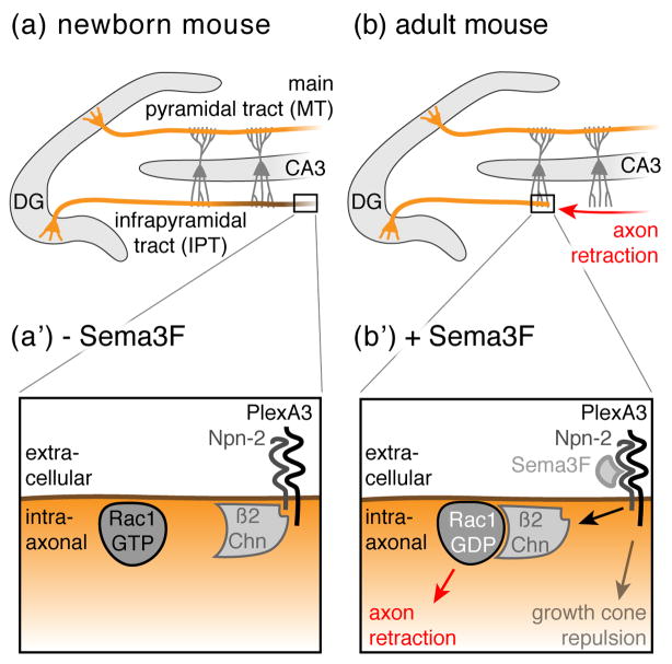
Figure 2. Axon retraction in the Dentate Gyrus.
(a) In neonatal mice, granule neurons (orange) located in the Dentate Gyrus (DG) project their axons (so called mossy fibers) into both the main and the infrapyramidal tracts (MT and IPT, respectively) to terminate on CA3 pyramidal dendrites (dark grey). (b) After two months, the IPT is remodeled through stereotyped retraction (red arrow) to generate adult structures. (a’ & b’) At the molecular level, Sema3F expression correlates with the regressive time period: in neonatal mice, Sema3F is not detectable (− Sema3F), however, shortly after birth, Sema3F expression is upregulated (+ Sema3F). Sema3F binds to the Neuropilin-2 (Npn-2) receptor, which in turn releases β2-Chimaerin (β2Chn) to axonal membranes where it triggers the hydrolysis of GTP to GTP + Pi in Rac1, thereby leading to axon retraction. By contrast, growth cone repulsion occurs independently of β2Chn.

