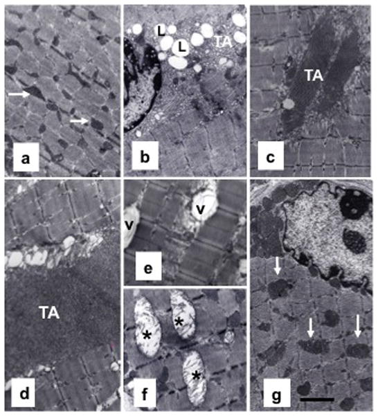
Fig. 2.

ØY+T: O joining improves myofibrillar ultrastructure in old mice (a–g). Representative TEM image of a portion of a gastrocnemius muscle from a young mouse exhibits normal myofibrillar cytoarchitecture and sarcomere organization (a) with abundance of mitochondria (arrow). In striking contrast, muscle from an old mouse exhibits perturbed muscle ultrastructure (b), including IML accumulation (L) and formation of large areas of tubular aggregation (TA). Muscle from an aged parabiont in Y: O pairing shows no IML accumulation and relatively smaller areas of TA formation (c). In contrast, muscles from aged partners in ØY: O pairing (d–f) show larger areas of TA formation and vacuolated (V) and swollen mitochondria with broken cristae (asterisk). Notably, ØY+T: O pairing restores normal cytoarchitecture and sarcomere organization with abundant hypertrophied mitochondria in aged mice (g). Scale bar = 2.5 μm.
