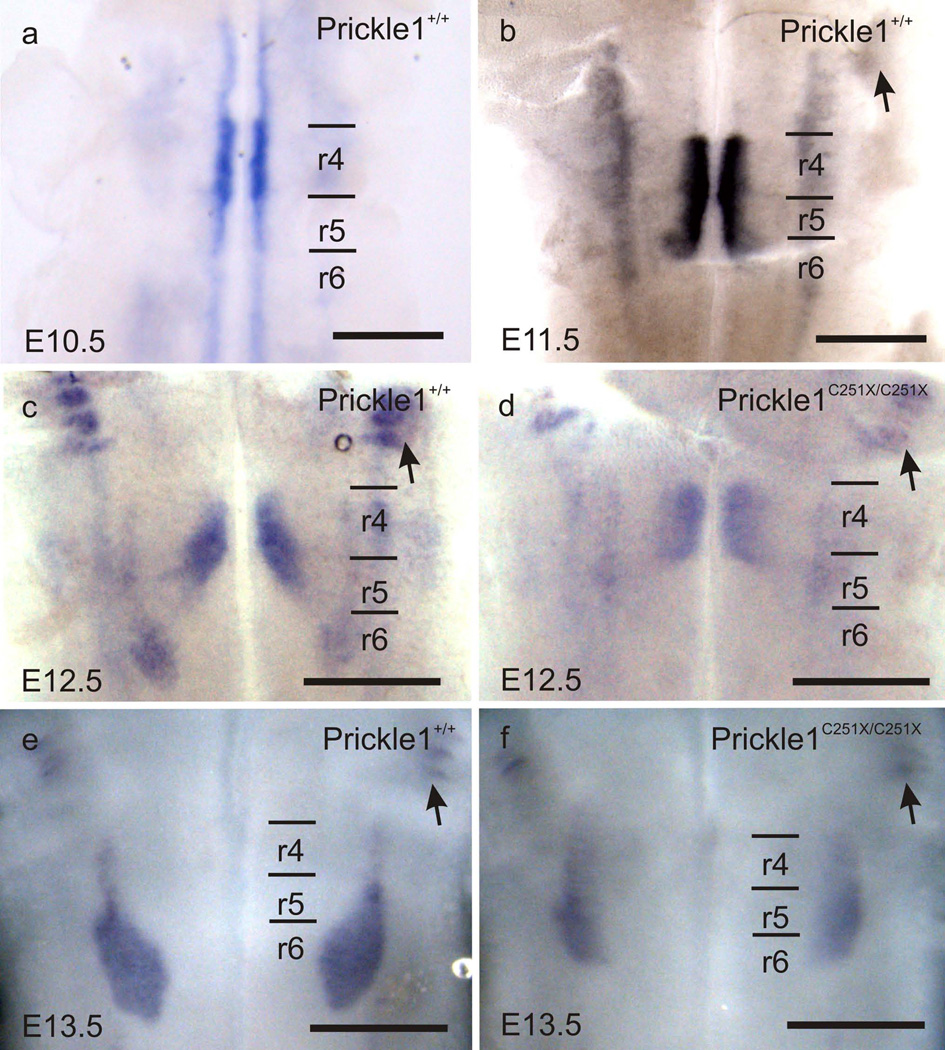
Fig. 1.
Prickle1 is expressed by the migrating FBMs as revealed by mRNA in situ hybridization. a: Prickle1 is highly expressed by the pre-migration FBMs at E10.5. In addition, Prickle1 expression is also detected in other motor neurons. b–f: Prickle1 is expressed by the FBMs from E11.5 to E13.5, and trigeminal neurons (arrows). d and f: The expression level in Prickle1C251X/C251X is reduced and the facial nucleus is barely visible. The FBM nucleus forms in r6 in the wild-type, but spans from r4 to r6 in the homozygotic mutant. Arrow: trigeminal neurons; r4–r6: rhombomere 4–6.The scale bar is 500µm

