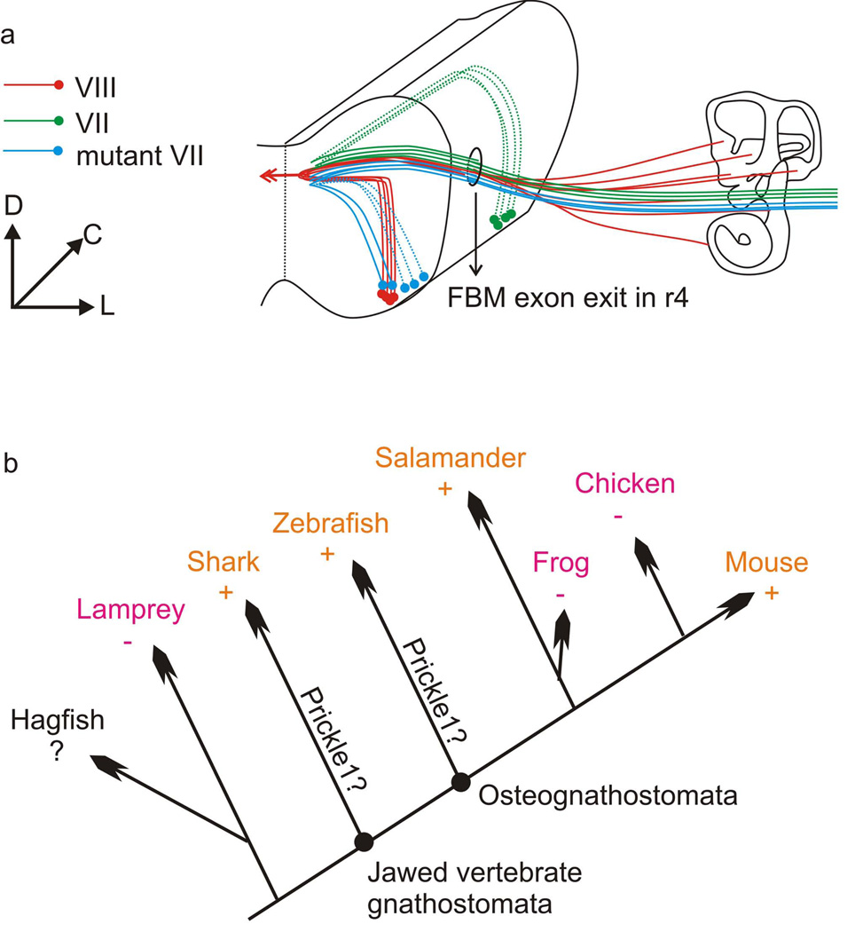
Fig. 7.
a: Diagram showing the migratory route of wild-type and Prickle1C251X/C251X FBMs. Both the olivo-cochlear efferents (VIII, red) and the facial nerve (VII, green) exit at r4. While the wild-type FBMs migrate caudally to r6, the mutant FBMs (blue) stay in r4 and r5 and lie dorsally to the olivo-cochlear efferent nucleus. b: Diagram showing the distribution FBM caudal migration (+) or lack of caudal migration (−) in vertebrates. Orange color highlights the presence of FBM caudal migration in some members, magenta color highlights the absence of FBM caudal migration in all members of a given lineage. Similarities of molecular data on FBM migration in mammals and zebrafish suggest that the common ancestor had such migration. This implies that frogs and birds have independently lost caudal FBM migration. C: caudal; D: dorsal; L: lateral; +: FBM migrates caudally; −: FBM does not migrate caudally; ?: data not available; Prickle1?: further analysis is required to show whether Prickle1 was present in this common ancestor and involved in FBM migration

