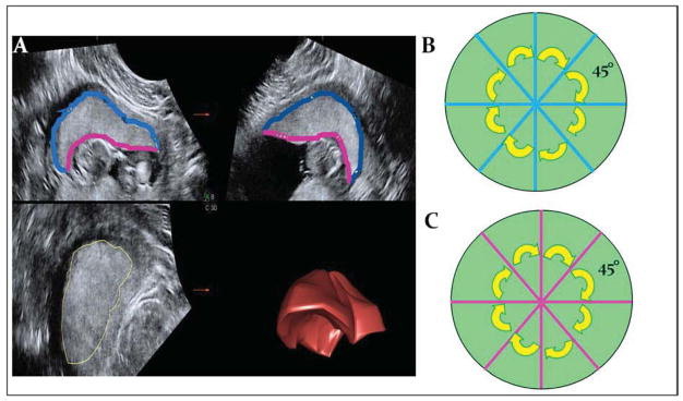
Figure 1. Measurement of the placenta using 3-dimensional ultrasonography.

1A shows the sectional plane display of the placental volume set, with quadrants A, B, and C corresponding to the 3 orthogonal planes. Successive tracings in plane A are rendered as a 3-dimensional placental volume in the lower right quadrant (3D). The maternal surface of the placenta is outlines in blue, while the fetal, or chorionic, surface is in pink. When the maternal (blue) and chorionic (pink) surfaces are measured in the A and B planes before and after a 45° rotation around the y-axis, this results in 4 evenly spaced measurements of each surface as depicted in 1B.
