Figure 1.
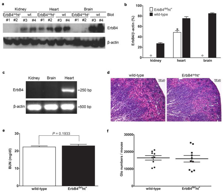
ErbB4 deletion in the kidneys of ErbB4delht+ mice. (a) ErbB4 deletion in ErbB4delht+ mice was confirmed by Western blots using an anti-ErbB4 antibody. wt: wild-type. (b) Densitometric analysis of the Western blot bands of ErbB4 in (a) and expressed as ErbB4/β-actin. *P < 0.05, ϕP < 0.001 compared to wild-type. (c) Heart excluded ErbB4 mRNA deletion was confirmed by RT-PCR. β-actin mRNA was used as a positive control. (d) Representative H&E staining of P10 kidneys from wild-type and ErbB4delht+ mice. n = 10 in each group. No apparent tubular dilatation or cyst formation was detected. (e) No significant difference of BUN levels between wild-type and ErbB4delht+ mice. n = 10 to 15 in each group. (f) Glomerular (Glo) numbers were calculated on adult mice and no significant differences were detected between wild-type and ErbB4delht+ mice. n = 9 to 10 in each group.

