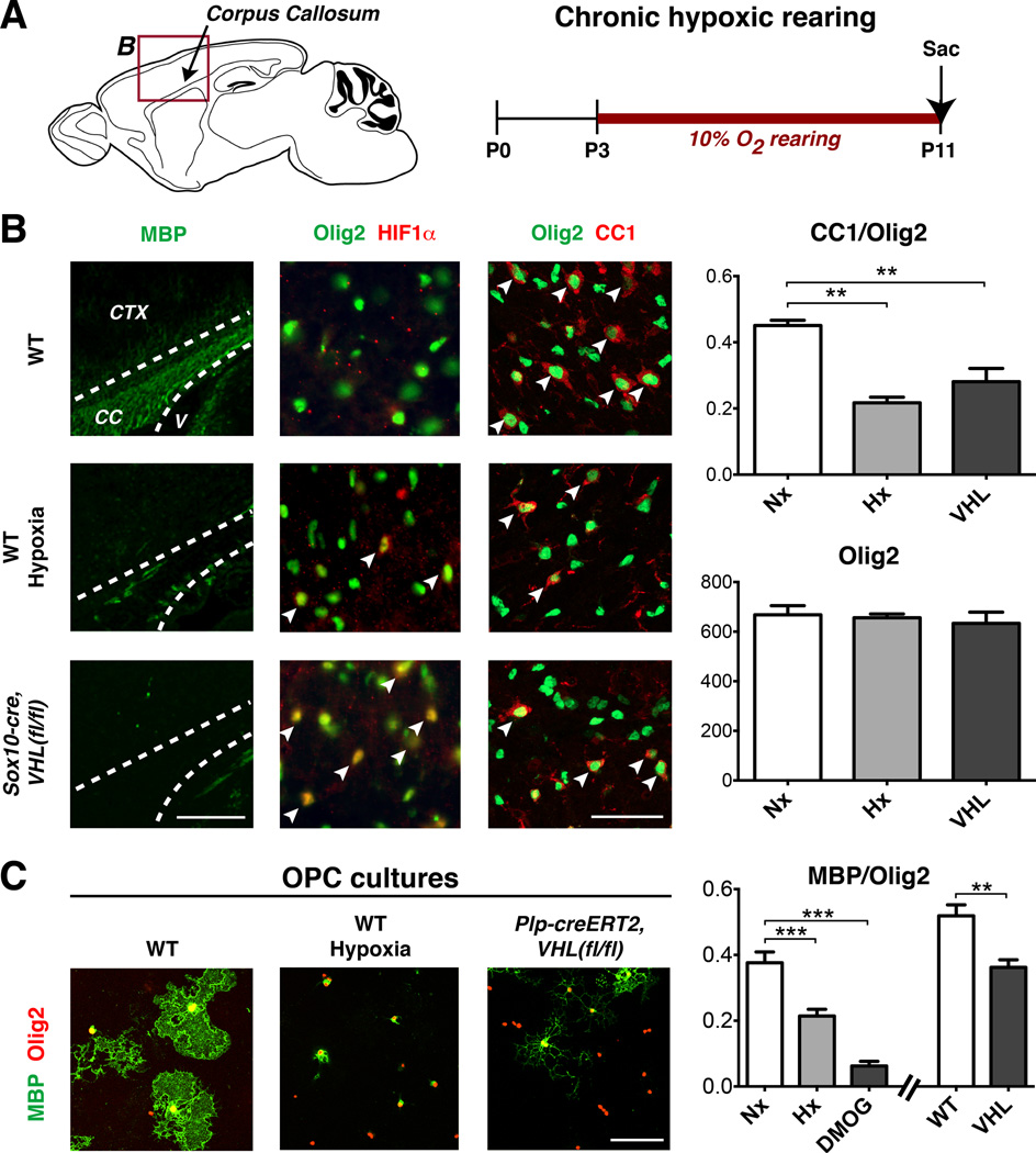
Figure 1. Oligodendrocyte-specific VHL deletion inhibits differentiation and myelination.
(A) Schematic of anatomical regions of corpus callosum (CC), cerebral cortex (CTX), and ventricle (V) presented in (B) and experimental timeline for chronic hypoxic rearing.
(B) Images showing hypomyelination, OL-lineage HIF1α expression, and OPC maturation arrest in CC of hypoxic WT mice or normoxic Sox10-cre, VHL(fl/fl) mice at P11. Arrowheads denote double-positive cells. Scale bar: 100µm (MBP), 50µm (Olig2).
(C) Immunopurified OPCs exposed to hypoxia or isolated from Plp-creERT2, VHL(fl/fl) mice show differentiation block. Scale bar: 100µm.
(For quantifications, mean+SEM; n≥3 experiments/genotype; **p<0.01, ***p<0.001; one-way ANOVA with Dunnett’s multiple comparison test)
See also Figure S1.

