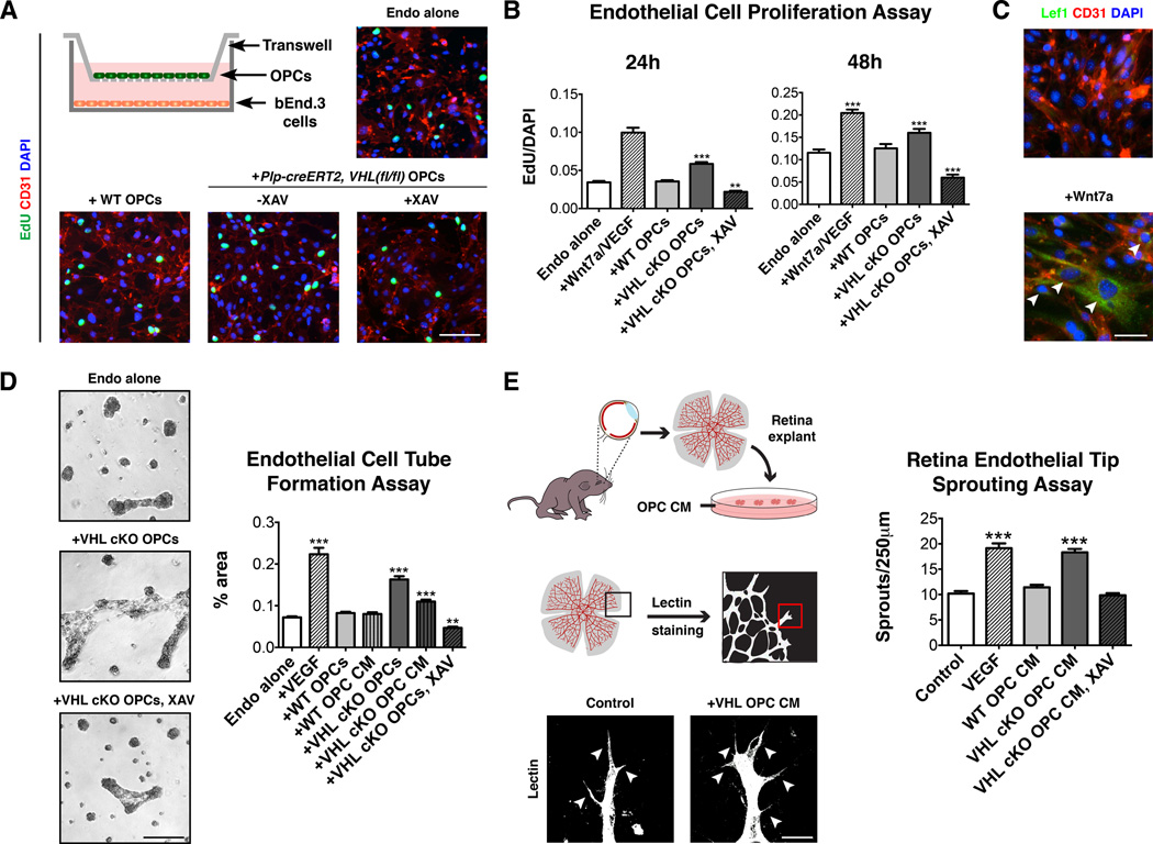
Figure 5. OPCs directly promote angiogenesis in a Wnt-dependent manner.
(A) Scheme showing transwell co-culture assay for OPCs and bEND.3 cells. OPCs from Plp-creERT2, VHL(fl/fl) mice induce endothelial cell proliferation in a Wnt-dependent manner. Scale bar: 100µm.
(B) Quantification of endothelial cell proliferation in transwell assay at 24h and 48h.
(C) Wnt7a treatment of bEND.3 cells induces Lef1 expression (arrowheads). Scale bar: 30µm.
(D) Transwell co-cultures of Plp-creERT2, VHL(fl/fl) OPCs and bEND.3 cells promotes endothelial cell tube formation in a Wnt-dependent manner. Scale bar: 500µm.
(E) Schematic showing retina endothelial tip sprouting assay. Conditioned medium from Plp-creERT2, VHL(fl/fl) OPCs promoted endothelial tip sprouting and filopodia extension in a Wnt-dependent manner. Scale bar: 25µm.
(For all quantifications mean+SEM; n≥3 experiments (A,B), n≥2 (D,E); **p<0.01, ***p<0.001; one-way ANOVA with Dunnett’s multiple comparison test)
See also Figure S5.

