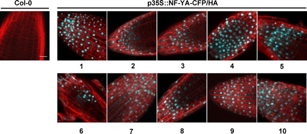
Fig. 3.

NF-YA proteins are nuclear-localized. Protein localization in Col-0 and p35S::NF-YA-CFP/HA overexpression lines (numbers below pictures represent the individual NF-YA genes). The cyan fluorescence protein (CFP) signal (blue) was always strongly associated with the nucleus (note that localization was confirmed by merged images, combining the CFP localization of NF-YAs with DIC imaging and Hoechst 33342 labeling staining of the nucleus—Fig. S6). The cell walls are stained with propidium iodide (red). The scale bar in Col-0 equals 15 μm
