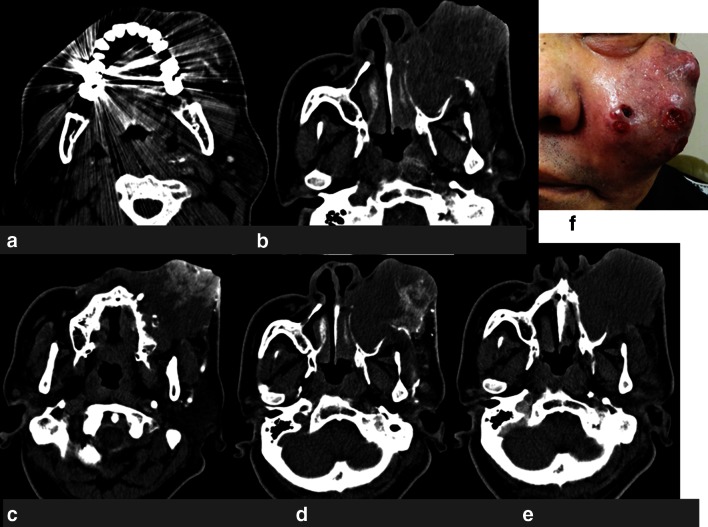
Fig. 1.
Case 1: a 66-year-old man with maxillary cancer (T4AN2bM0). a CT angiography obtained in the selected left side maxillary artery. It was difficult to confirm the vascular territory due to dental metals. b CT angiography obtained in the selected left side maxillary artery. It was difficult to confirm the vascular territory due to obstacle enhancement. c CT angiography obtained in the selected left side facial artery. It was sufficiently clear to confirm the vascular territory. d CT angiography obtained in the selected left side transverse facial artery. It was sufficiently clear to confirm the vascular territory. e CT angiography obtained in the selected left side internal carotid artery. It was difficult to confirm the vascular territory due to obstacle enhancement. f Maxillary cancer invading the face before treatment

