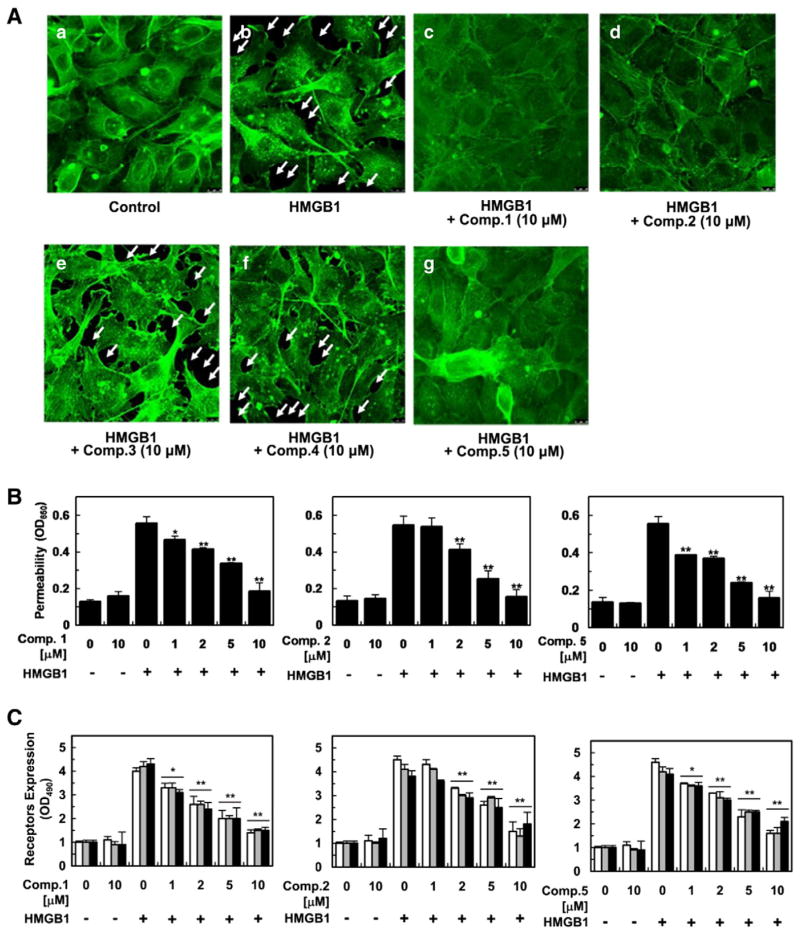
Fig. 4.
In vitro evaluation of vascular protective action of compounds 1, 2, and 5 against HMGB1 (1 μg/mL for 16 h)-induced vascular barrier disruption. Data were analyzed upon pretreatment of HUVECs with the indicated concentrations for 6 h. Staining for F-actin upon pretreatment of 1–5 (A). HMGB1-induced permeability was monitored by assessing the flux of Evans blue bound albumin across HUVECs (B). Influence of 1, 2, and 5 on expression of HMGB1 receptors (C), phosphorylation of p38 (D), and CAMs (E)was evaluated by ELISA. White, gray, and black bars indicate expression of TLR2, TLR4, and RAGE (C) and VCAM-1, ICAM-1, and E-selectin, respectively (E). Cell adhesion inhibitory action against enhanced cell-cell adhesion by HMGB1-induced vascular stress was assessed utilizing fluorescently labeled THP-1 cells (F). Biological results of inactive compounds (3, 4) are shown in Supplementary data 2. Results are indicated with the means ± SDs of different of at least three experiments and *p < 0.05 and **p < 0.01 compared to cells only treated with HMGB1 (B–F).


