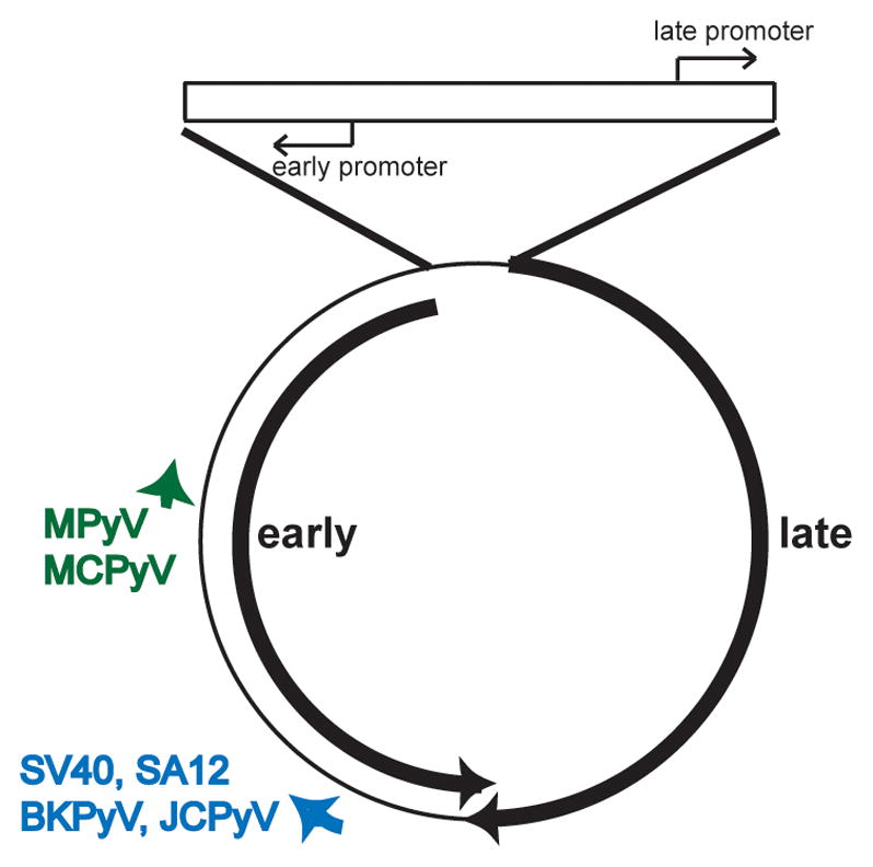
Figure 1.

Map of the polyomavirus genome. The double stranded, circular DNA genome of the polyomaviruses described in the text is shown along with the regulatory region (top), and early and late primary RNA transcripts (black heavy arrows). The positions of the miRNAs (short heavy arrows) are also indicated. The miRNAs are expressed from the same strand as the late transcripts and are therefore perfectly complementary to the early mRNAs. As such, they target the early mRNAs for degradation. In BPCV, the miRNA maps to an additional non-coding region between the early and late transcripts
