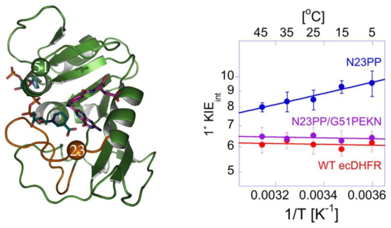
Figure 4. Evolutionary Preservation of DHFR Dynamics.

Left Panel: Structure of WT DHFR showing the positions of N23 and G51. The Met-20 loop is shown in brown, folate is shown in purple, and the nicotinamide ring of NADPH is in blue. Residues N23 and G51 are shown as brown and green spheres, respectively. Right Panel: Arrhenius plots for the intrinsic H/T KIEs for the WT, N23PP, N23PP/G51PEKN [1]. Reprinted with permission from the American Society of Biochemistry and Molecular Biology.
