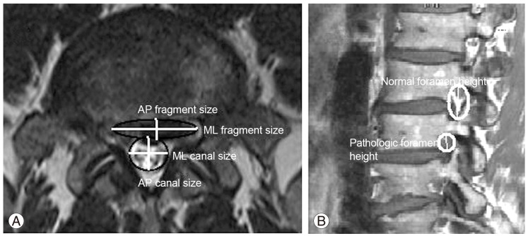
Fig. 1.
(A) Axial magnetic resonance imaging in a patient with a protruded disc. The disc fragment and thecal sac are schematically shown as an oval and circle, respectively, and their diameters as AP and ML sizes. The ratios are calculated as: AP fragment ratio=AP fragment size/AP canal size; ML fragment ratio=ML fragment size/ML canal size. (B) A sagittal magnetic resonance imaging in a different patient. Neural foramens are schematically shown as ovals encircling peri-neural fat. Foramen ratio=pathologic foramen height/normal foramen height. AP, anteroposterior; ML, mediolateral.

