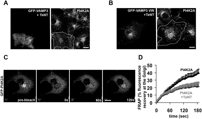
Fig. 2.
PI4K2A localization is affected by VAMP3 inactivation. (A,B) Fluorescence micrographs of COS-7 cells co-transfected with TeNT and either GFP–VAMP3 wt (A) or the GFP–VAMP3 VW mutant (B). Fixed cells were stained with a rabbit polyclonal antibody against PI4K2A (right panels). The dotted line indicates the cell outline. (C,D) Retrograde transport of PI4K2A following photobleaching of the perinuclear and Golgi compartment in COS-7 cells expressing GFP–PI4K2A. (C) Sequential imaging during photobleaching. (D) The rate of FRAP was faster in cells transfected with GFP–PI4K2A alone (black, n = 10), than in those coexpressing GFP–PI4K2A and TeNT (gray, n = 10). Shown are mean fluorescence intensity curves +s.e.m. (PI4K2A alone) or –s.e.m. (PI4K2A + TeNT), plotted as described in the Materials and Methods. Scale bars: 10 µm.

