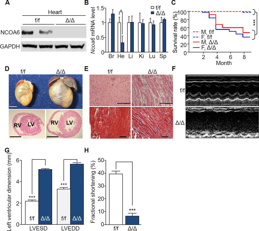
Figure 1. Premature death and impaired cardiac function in cardiomyocyte-specific Ncoa6 knockout mice.
(A) Western blot analysis of NCOA6 in the hearts of one-month-old f/f and Δ/Δ male mice. GAPDH was used as a loading control.
(B) RT-qPCR analysis of Ncoa6 mRNA in various organs of one-month-old f/f and Δ/Δ male mice. Br, Brain; He, Heart; Li, Liver; Ki, Kidney; Lu, Lung; Sp, Spleen. Ncoa6 mRNA levels were normalized to those of Gapdh. Graphs show means ± s.d.; *P < 0.05.
(C) Kaplan-Meier survival curves of f/f (male, n = 34; female, n = 34) and Δ/Δ (male, n = 21; female, n = 39) mice. Genders and genotypes are labeled inside the plot. M, male; F, female. ***P < 0.001.
(D) Gross morphology (top) and histological examinations (bottom, H&E) of hearts from 4-month-old female f/f and Δ/Δ mice. LV, left ventricle; RV, right ventricle. Scale bars, 2.5 mm.
(E) H&E (top) and Masson’s trichrome staining (bottom) of left ventricles from 4-month-old female mice. Scale bars, 100 ìm.
(F–H) Representative profiles of M-mode echocardiographic analyses (F) and quantitative representations of LVESD and LVEDD (G) and percent fractional shortening (H) of f/f (male, n = 3; female, n = 4), and Δ/Δ (male, n = 3; female, n = 3) mice. Graphs show means ± s.d.; ***P < 0.001. See also Figure S1 and S2.

