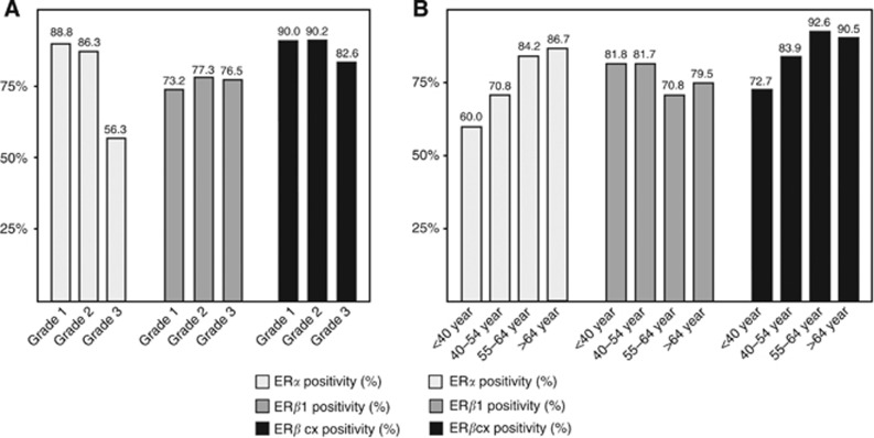
Figure 1.
ER expression among different age groups and histological grades. (A) ERα positivity decreased with higher Elston–Ellis histological grade (white; P<0.001, N=80, 168, and 71). ERβcx showed a similar trend, however not significant (black; P=0.23, N=80, 163, and 69). ERβ1 was equally distributed among all three grades (grey; P=0.771, N=82, 163, and 68). (B) Patients were divided into four different age groups according to age at diagnosis, <40, 40–54, 55–64, and ⩾65 years. ERα (white) positivity was lowest in the youngest age group and increased with age (P=0.011, N=10, 96, 139, and 75). A similar trend was seen with ERβcx (black), however not reaching significance (P=0.062, N=11, 93, 135, and 73). ERβ1 was equally distributed along all age groups (grey; P=0.221, N=11, 93, 137, and 74). Numbers above bars reflect percentage of positive tumours.

