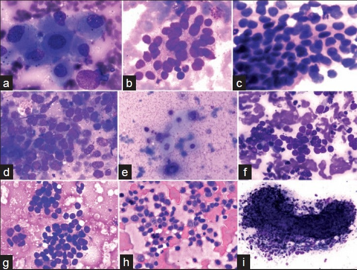
Figure 1.

(a) A cluster of large pleomorphic cells with abundant cytoplasm, vesicular nuclei and prominent nucleoli in an aspirate from a case of hepatocellular carcinoma (MGG, ×400). (b) Mucus secreting adenocarcinomatous metastasis showing a loose cluster of markedly pleomorphic vesicular cells with abundant cytoplasm and indistinct cell borders (MGG, ×00). (c) Metastatic ductal carcinoma breast showing cohesive cell cluster. Nuclei are vesicular and overlapping (H and E, ×400). (d) A cohesive cluster of mildly pleomorphic hyperchromatic adenocarcinomatous cells with minimal cytoplasm from a case of gallbladder adenosquamous carcinoma (MGG, ×400). (e) Malignant squamous cells from the above case have hyperchromatic nuclei and abundant glassy-blue cytoplasm (MGG, ×400). (f) Metastatic small cell anaplastic carcinoma from lung showing a cohesive cluster of hyperchromatic cells; nuclear molding can be appreciated (MGG, ×400). (g) Metastatic malignant round cell tumor showing a cluster of round-ovoid cells with scanty cytoplasm. Rosettes are evident (MGG, ×400). (h) Infiltration of Non-Hodgkins lymphoma cells: Discretely present large cells have irregular nuclear membrane and prominent nucleoli (H and E, ×400). (i) Metastatic spindle cell sarcoma showing a “microbiopsy” of ovoid — spindle cells; discrete cells present at the periphery (MGG, ×100)
