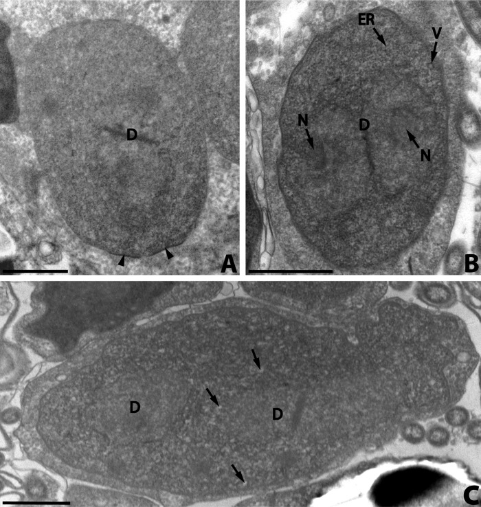
Figure 2.
Nosema podocotyloidis n. sp. A. Young sporont showing the thickening of the wall (arrowheads). B. Sporont showing electron-lucent vesicles (V) and a few rough endoplasmic reticula (ER). C. Sporont with two diplokarya (D). Electron-lucent vesicles (arrows). D: diplokaryon; ER: endoplasmic reticulum; N: nucleolus-like formation; V: electron-lucent vesicles. Scale Bars: A, B, C and D, 1 μm.

