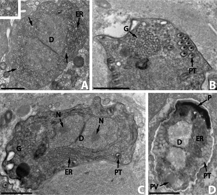
Figure 3.
Nosema podocotyloidis n. sp. A. Young sporoblast showing numerous electron-lucent vesicles (arrows and insert) and the endoplasmic reticulum (ER). B. A part of the sporoblast showing the polar tube (PT) and the Golgi apparatus (G). C. Elongate sporoblast. Note the presence of the Golgi apparatus (G). D. Immature spore with 12 coils of polar tube. D: diplokaryon; ER: endoplasmic reticulum; N: nucleolus; P: polaroplast; PT: polar tube; PV: posterior vacuole. Scale Bars: A, B, C and D, 1 μm.

