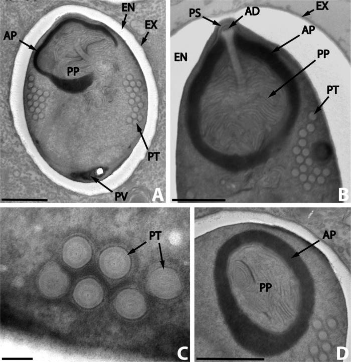
Figure 4.
Nosema podocotyloidis n. sp. A. Mature spore with 15 coils of polar tube. B. Detail of the anterior region of a mature spore showing the anchoring disc (AD) and the polar sac (PS). C. Transversally sectioned polar tube coils revealing the different layers. D. Detail of the polaroplast revealing the structure of the outer or anterior region (AP) and of the inner or posterior region (PP). AP: anterior polaroplast; EN: endospore; EX: exospore; PT: polar tube; PP: posterior polaroplast; PS: polar sac; PV: posterior vacuole. Scale Bars: A, B and D, 1 μm; C, 0.1 μm.

