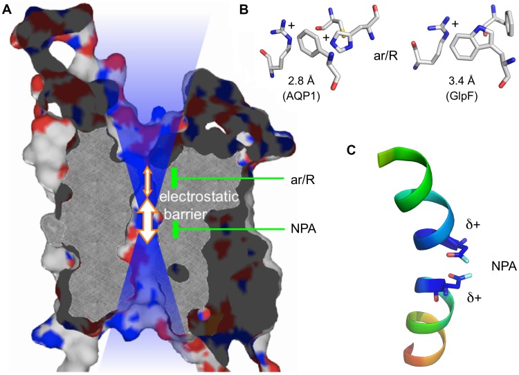
FIGURE 1.
Protein structure of an AQP monomer. (A) Slab of AQP1 showing the channel and the two constriction sites. The positive electrostatic field emanating from the channel is symbolized by the blue transparent shading. (B) Typical amino acid composition of the ar/R region in a water specific AQP (here AQP1; PDB #1J4N; Borgnia et al., 1999) and an aquaglyceroporin (here Escherichia coli GlpF; PDB #1FX8; Fu et al., 2000). (C) Half helices B and E with the capping asparagine residues residing at the positive helix ends.

