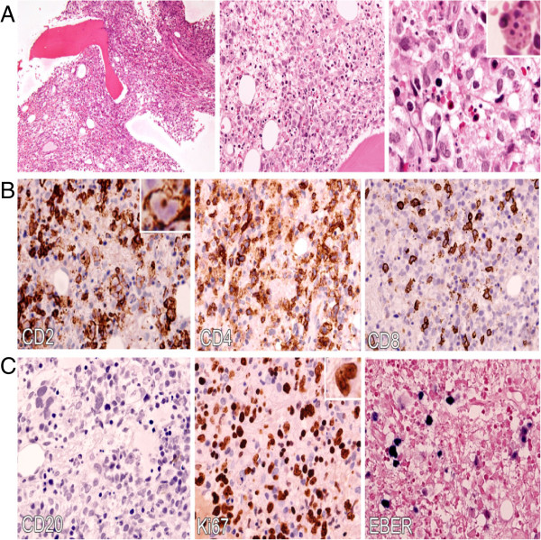
Figure 1.

Systemic Epstein-Barr virus-positive T-cell lymphoproliferative childhood disease: bone marrow biopsy. A: Hematoxylin and eosin (H&E) sections illustrate the medium-sized lymphoid cells replacing the normal hematopoietic elements and exhibiting moderate atypia with irregular kidney-shaped nuclei, dispersed chromatin and distinct nucleoli (100x, 400x and 600x respectively left to right). Hemophagocytosis is also easily seen (inset). B: An immunohistochemical study shows that the majority of infiltrated atypical lymphocytes expressed CD2+ and CD4+ with scattered positivity to CD8. C. The atypical lymphoid cells are not immunoreactive to CD20 and there is a paucity of B cells in the bone marrow. Ki-67 immunostaining shows a high proliferative index and EBV-encoded RNA (EBER) by in situ hybridization demonstrates a strong reactivity of atypical lymphoid cells.
