
Figure 3.
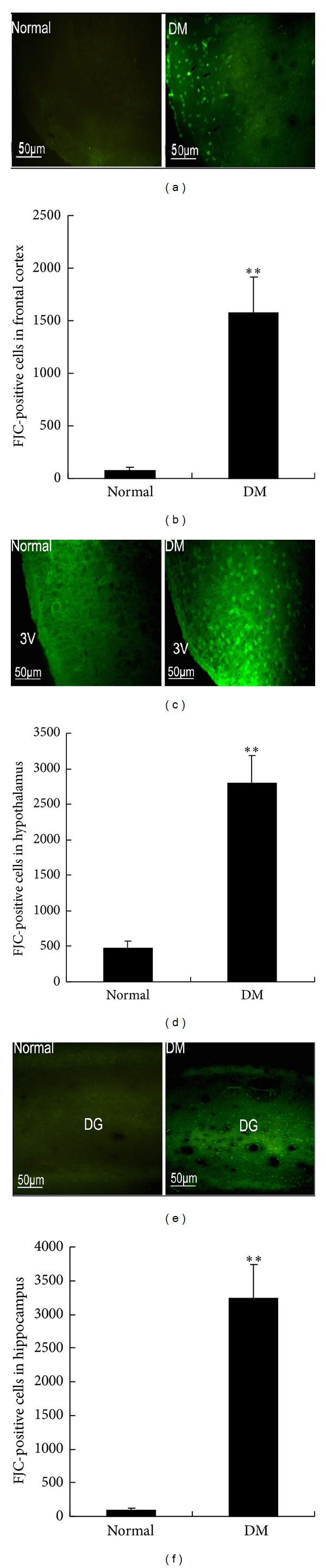
Fluoro-Jade C- (FJC-) positive cells were examined in the frontal cortex, hypothalamus, and hippocampus of rats at 4 months after STZ injection. (a) Representative images of FJC-positive cells in the frontal cortex. (c) Representative images of FJC-positive cells in the hypothalamus; 3V: third ventricle. (e) Representative images of FJC-positive cells in the hippocampus; DG: dentate gyrus. (b) Frontal cortex, (d) hypothalamus, and (f) hippocampus. FJC-positive cells were counted (see Materials and Methods for procedure details). **P < 0.01, significant difference compared with the age-matched control rats; n = 6.