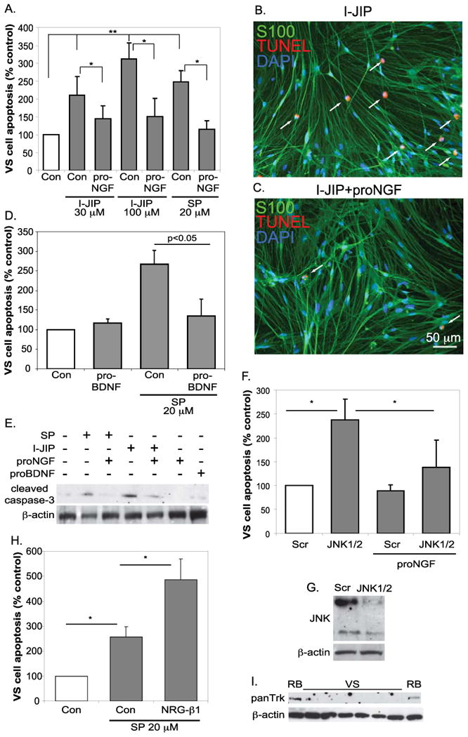
Figure 4.

ProNGF protects VS cells from apoptosis due to JNK inhibition. A-D. Primary VS cultures were maintained in the presence of the JNK inhibitors, I-JIP or SP600125, with or without treatment with proNGF (3 nM) (A) of proBDNF (3 nM) (D). Cultures were immunostained with anti-S100 (green) and labeled with TUNEL (red). Nuclei were labeled with DAPI (blue). The average percent of TUNEL-positive, S100-positive condensed nuclei were scored and plotted relative to cultures not treated with JNK inhibitors or proNGF (control). Data are from cultures derived from six separate patients. *p<0.05, **p<0.01 by one way ANOVA followed by Dunn's method. E. Western blot of protein lysates from primary VS cultures treated with the indicated reagents probed with anti-cleaved caspase-3 antibodies. Blots were stripped and reprobed with anti-β-actin antibodies. F. Average percent apoptosis in primary VS cultures transfected with scrambled or anti-JNK1/2 siRNA oligonucleotides and maintained in the presence or absence of proNGF. Comparison for differences among conditions by one way ANOVA followed by Dunn's method. G. Western blot of protein lysates from primary VS cultures transfected with scrambled (Scr) or JNK1/2 targeted siRNA oligonucleotides and probed with anti-JNK antibodies. Blots were stripped and reprobed with anti-b-actin antibodies. H. Average percent apoptosis in primary VS cultures treated with or without neuregulin (NRG) β-1 and maintained in the presence or absence of proNGF. Comparison for differences among conditions by Kruskal-Wallis one way ANOVA on ranks. I. Western blot of protein lysates from primary VS and rat brain (RB) specimens with an anti-panTrk antibody. The blots were stripped and reprobed with anti-β actin antibodies.
