
Fig. 1.
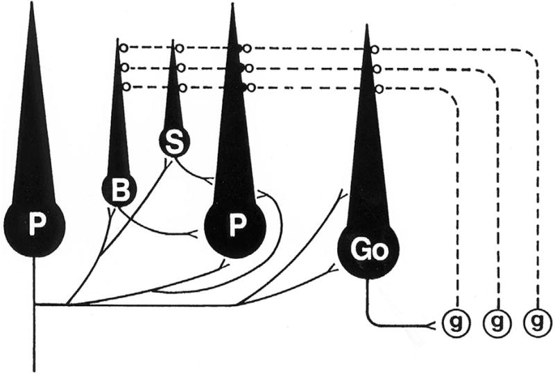
Circuit diagram of the major cerebellar cortical neurons and their projections. Absent from the diagram are the extracerebellar afferents, as would be the case in an isolated cerebellar culture. Projecting axons from Purkinje cells are shown in the Purkinje cell on the left side of the diagram, while projections to Purkinje cells from other neurons are shown in the Purkinje cell in the center. Both Purkinje cells would, of course, have both afferent and efferent projections. The only excitatory cortical neurons are the granule cells (g), and their axons, the parallel fibers (shown as dashed lines), project to the dendrites of all other cortical neurons. All other cortical neurons are inhibitory, and their axons are represented as solid lines. Purkinje cells (P) are the only neurons whose axons project from the cortex, predominantly to the deep cerebellar nuclei (not shown). Purkinje cell axon collaterals project to all other inhibitory cortical neurons, including other Purkinje cells. Basket cell axons (B) project to Purkinje cell somata and proximal dendrites, while stellate cell (S) axons inhibit more distal parts of the Purkinje cell dendritic tree. Purkinje cell somata thus receive inhibitory projections from only two cortical sources, namely basket cells and recurrent axon collaterals from other Purkinje cells. Golgi cells (Go) project their complex axons to granule cell dendrites. (From Seil, 1996, with permission).