
Fig. 2.
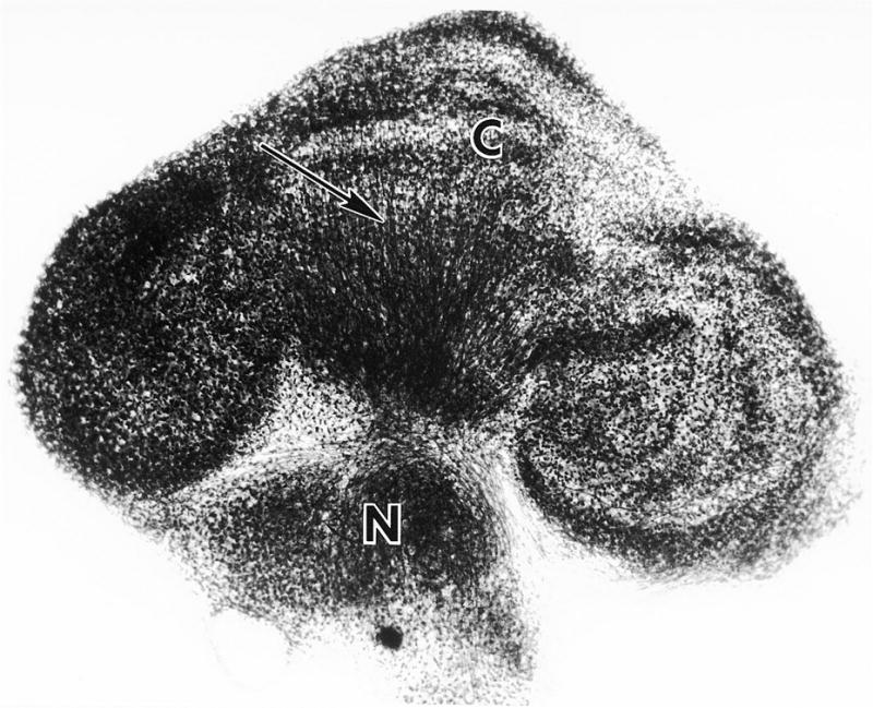
Low power view of a newborn mouse derived cerebellar culture after 23 DIV. The explant is oriented in the parasagittal plane. The cortical region (C) is readily distinguishable from an incorporated group of deep cerebellar nucleus neurons (N). Laminae are evident in regions of the cortex. Axons (arrow) of Purkinje cell origin project to the deep nucleus. The gross anatomical relationships are similar to those in the cerebellum in vivo. Whole mount preparation, Holmes silver stain, X75. From Seil and Leiman, 1977, with permission).