
Figure 1. CCN2 mRNA and protein are present in mouse HSC exosomes.
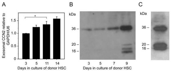
(A) Quantitative RT-PCR of CCN2 mRNA in exosomes isolated from primary mouse HSC on Day 3-14 of culture. Data are from three independent experiments and expressed as mean ± s.e.m. *P<0.05 as determined by Student’s t-test using SIGMA PLOT 11.0 software (SPSS Inc., Chicago, IL). (B) Western blot for CCN2 in exosomes isolated from 24-hour conditioned medium from individual T-75 flasks containing primary mouse HSC on days 3-9 of culture (5 g total exosomal protein/lane). (C) CCN2 Western blot of exosomes isolated from P5 mouse HSC. Data are representative of three independent determinations.