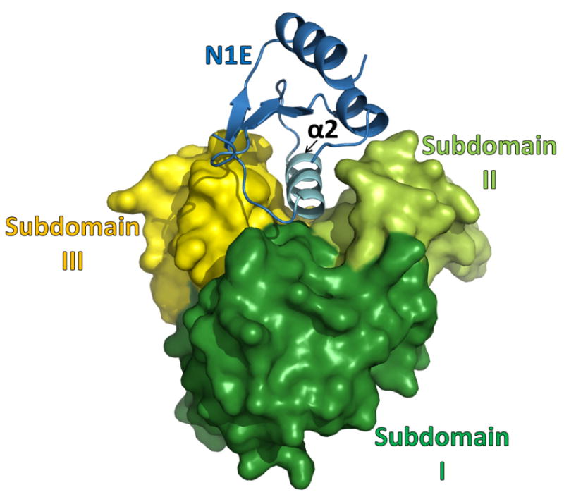
Fig. 3. Structure of the V. vulnificus N1E•cyto-GspL heterodimer.

The N1E of GspE shown in blue, subdomain I of cyto-GspL in green, subdomain II of cyto-GspL in lime, subdomain III of cyto-GspL in yellow. Note how all three GspL subdomains are involved in contacting N1E with helix α2 of N1E, a major component of the interface. When compared with the V. cholerae N1E•cyto-GspL heterodimer (Abendroth et al., 2005) the r.m.s.d. is 1.4 Å with a difference of ∼11 degrees in N1E-vs-cyto-GspL orientation, and ∼48 and 53 % amino acid sequence identity for N1E and cyto-GspL, respectively (see also Supplementary Fig. S2 and S3).
