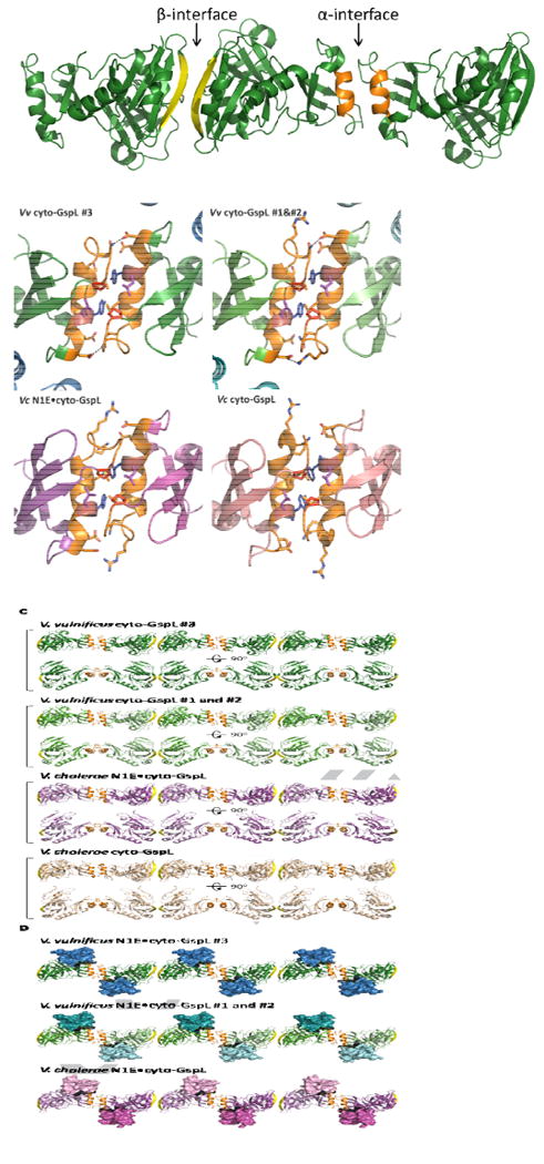
Fig. 5. Linear arrangements of Vibrio cyto-GspL domains in multiple crystals.

A. The α- and β-interfaces among cyto-GspL domains in the current V. vulnificus GspE•cyto-GspL crystals. The interfaces between cyto-GspL domain #3 and crystallographically related domains are depicted. The interfaces between cyto-GspL domains #1 and #2 are essentially the same (see text). The α-interface is formed mainly by side chain contacts between residues of α2 helices (orange) and the loops between strand βE and helix α2 from two domains. The domains are related by a twofold axis approximately parallel to the direction of view. (See text and Fig. 5B for further description of the contacts). In the β-interface, main chain hydrogen bonds between antiparallel βc strands (yellow) are the main contacts between two subunits, which are also related by a twofold approximately parallel to the direction of view.
B. Close-ups of four similar cyto-GspL α-interfaces in three different crystal forms. Key residues are shown in sticks. Hydrogen bonds and electrostatic interactions are indicated with dashed lines. Note the completely conserved Pro70 (red) in all interfaces, twice in contact with the highly conserved Tyr83 (blue) and Leu84 (purple).
Left upper: The α-interface between two neighboring cyto-GspL domains #3 in the current V. vulnificus GspE•cyto-GspL crystals.
Right upper: The α-interface between neighboring cyto-GspL domains #1 and #2 in the current V. vulnificus GspE•cyto-GspL crystals.
Left lower: The α-interface between neighboring cyto-GspL domains in the V. cholerae N1E•cyto-GspL crystals (PDB 2BH1) (Abendroth et al., 2005).
Right lower: The α-interface between neighboring cyto-GspL domains in the V. cholerae cyto-GspL crystals (PDB 1YF5) (Abendroth et al., 2004a)
C. Four similar linear arrangements of cyto-GspL domains in three different crystal forms.
From top to bottom: cyto-GspL rods from: V. vulnificus GspE•cyto-GspL complex #3; V. vulnificus GspE•cyto-GspL complex#1 and #2; V. cholerae N1E•cyto-GspL; and V. cholerae cyto-GspL. The α-interface and β-interfaces are colored orange and yellow, respectively.
D. Three linear arrangements of cyto-GspL domains with associated N1E domains in two different crystal structures. From top to bottom: V. vulnificus GspE•cyto-GspL complex #3; V. vulnificus GspE•cyto-GspL complex #1 and #2; and V. cholerae N1E•cyto-GspL The α-interface and β-interfaces are colored orange and yellow, respectively. N1E domains are shown in surface representation.
