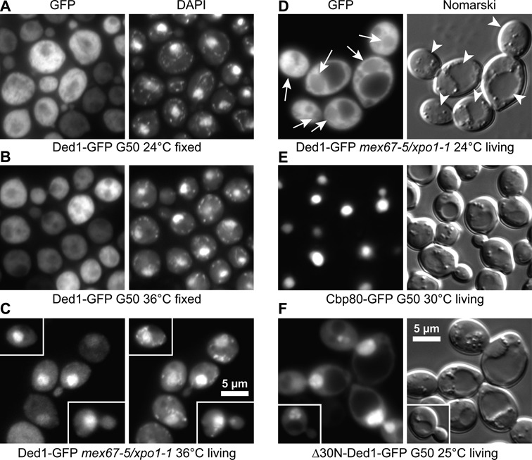
Figure 1.

Ded1 shuttled between the cytoplasm and nucleus. (A) and (B) Fluorescence microscopy of cells that were fixed with formaldehyde when stained with DAPI to reveal the nuclei and mitochondria (seen as speckles). Ded1-GFP was primarily located in the cytoplasm of wildtype (G50) cells at all the temperatures tested. (C) In contrast, Ded1-GFP shows a strong nuclear location in the mex67-5/xpo1-1 mutant when incubated at nonpermissive temperatures for 30 min in living cells stained with DAPI. (D) Fluorescence microscopy (GFP) and Nomarski interference contrast microscopy of living yeast cells. Ded1-GFP was mostly located in the cytoplasm of mex67-5/xpo1-1 cells at the permissive temperatures (24°C), but a small amount was visible in the nuclei as well (arrows). The vacuoles/lysosomes are indicated with arrowheads. (E) In contrast, Cbp80-GFP was almost exclusively located in the nuclei. (F) Ded1-GFP that was deleted for the 30 amino-terminal residues containing the NES sequence (Δ30N) and the eIF4E binding site accumulated in the nuclei of yeast even in wildtype cells.
