
Figure 5.
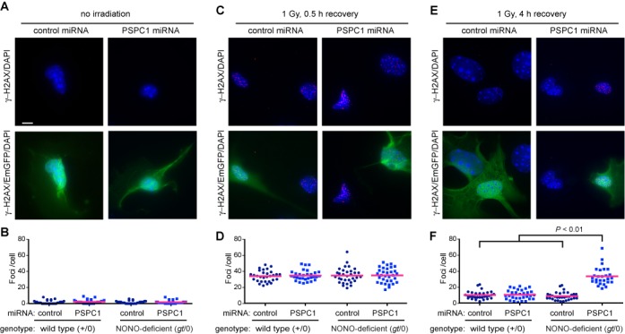
Delayed resolution of repair foci. Vectors encoding control or PSPC1 miRNA were introduced by electroporation into wild-type (+/0) or NONO-deficient (gt/0) MEFs. Cells were mock irradiated or exposed to 1 Gy of 137Cs γ-rays. At indicated times following irradiation, cells were fixed and analyzed by indirect immunofluorescence using anti-γ-H2AX primary and red secondary antibody and DAPI counterstain. (A, C, E) Representative fields showing γ-H2AX/DAPI channels (top row) or γ-H2AX/EmGFP/DAPI-merged images in NONO-deficient (gt/0) MEFs. Images were collected for non-irradiated control cells or for cells that were irradiated and allowed to recover for indicated times. Cell morphology and foci appearance were indistinguishable in wild-type (+/0) and NONO-deficient (gt/0) MEFs; only the latter are shown in the figure. Scale bar denotes 10 μm. (B, D, F) Foci per cell in populations corresponding to panels (A, C, E). A total of 30 nuclei were scored per experimental group. Blue symbols depict score for individual cells; red bar depicts mean. Treatment of NONO-deficient cells with PSPC1 RNA resulted in a significant increase in residual foci at 4 h post-irradiation (P < 0.01).