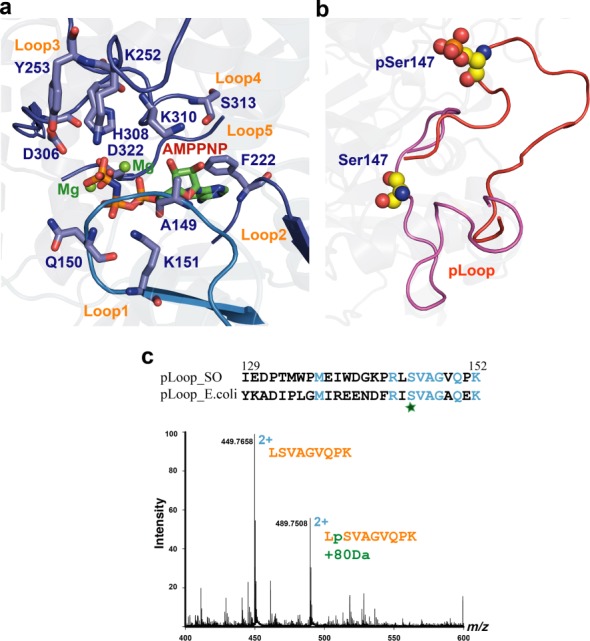
Figure 4.

S. oneidensis MR-1 HipAso is regulated conformationally upon autophosphorylation. (a) Structural detail of the AMPPMP (atom colored) and Mg2+ (green spheres) binding site in HipAso (blue). Residues involved are shown in stick representation. (b) Detailed view of the ejection of the pLoop of HipAso upon autophosphorylation. Ser147 and phosphate represented as spheres. (c) Identification of the phosphorylated peptide by nanoLCMS. The peptide LSVAGVQPK was observed at charge state 2+ in two forms differing by 80 Da in molecular weight. Sequence alignment of the pLoop regions in S. oneidensis MR-1 and E. coli.
