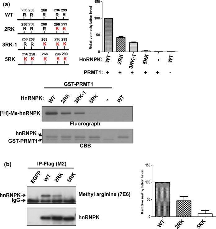
Figure 1.

Arg296 and Arg299 are the major methylation sites of hnRNPK in vitro and in vivo. (a) Detection and quantification of in vitro hnRNPK methylation levels and the arginine mutants (2RK, 3RK-1 and 5RK, as shown in the schematic diagram). HnRNPK and diverse mutants were incubated with GST-PRMT1 in the presence of [3H] S-adenosylmethionine (SAM), followed by SDS-PAGE. The proteins were stained with Coomassie blue (bottom). Methylation was detected through fluorography (top) and quantified using a liquid scintillation counter. The methylation levels of all arginine mutants relative to wild-type (WT) hnRNPK are shown as a bar graph. (b) Detection and quantification of in vivo methylation levels of WT hnRNPK and arginine mutants (2RK, 5RK). Flag-hnRNPKs were immunoprecipitated from the DF-1 cells expressing Flag-hnRNPKs using an anti-Flag antibody (M2). Protein expression was determined using an anti-hnRNPK antibody (bottom). Methylation levels were detected using an antibody against anti-asymmetric di-methylated arginine (top) and quantified using ImageJ software. The relative methylation levels of 2RK and 5RK mutants compared with WT hnRNPK are shown as a bar graph.
