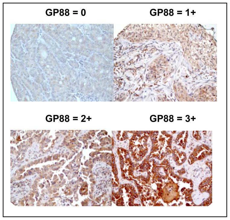
Figure 1. Immunohistochemical staining for GP88 expression in paraffin-embedded local NSCLC tissue sections.
Representative photomicrographs (magnification x200) of lung cancer tissue sections from local resected NSCLC showing various levels of GP88 expression with 0, 1+, 2+, and 3+ scores, respectively are provided.

