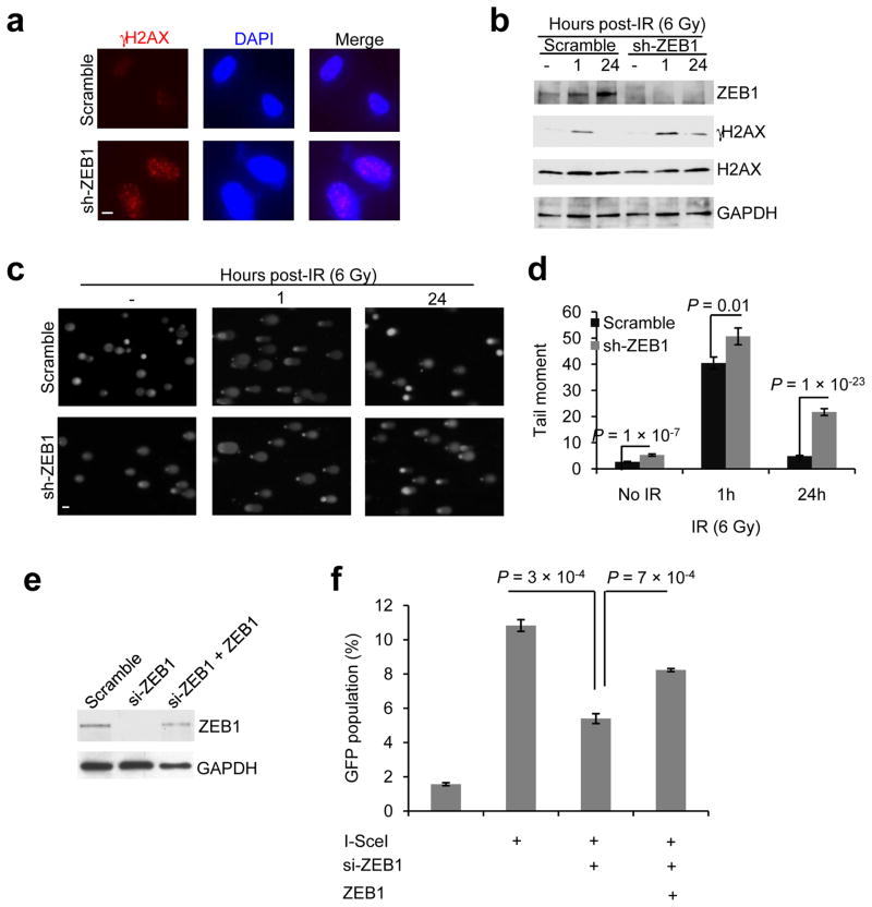
Figure 3. ZEB1 regulates DNA damage repair.
(a) γH2AX and DAPI staining of SUM159-P2 cells transduced with ZEB1 shRNA, 24 hr after 6 Gy IR. Scale bar: 10 μm.
(b) Immunoblotting of ZEB1, γH2AX, H2AX and GAPDH in SUM159-P2 cells transduced with ZEB1 shRNA, at the indicated time points after 6 Gy IR.
(c, d) Images (c) and data quantification (d) of comet assays of SUM159-P2 cells transduced with ZEB1 shRNA, at the indicated time points after 6 Gy IR. n = 62 cells per group. Scale bar in (c): 50 μm.
(e) Immunoblotting of ZEB1 and GAPDH in U2OS_DR-GFP cells transfected with ZEB1 siRNA alone or in combination with ZEB1.
(f) HR repair assays of U2OS_DR-GFP cells transfected with ZEB1 siRNA alone or in combination with ZEB1. n = 3 wells per group.
Data in d and f are the mean of biological replicates from a representative experiment, and error bars indicate s.e.m. Statistical significance was determined by a two-tailed, unpaired Student’s t-test. The experiments were repeated 3 times. The source data can be found in Supplementary Table 3. Uncropped images of blots are shown in Supplementary Figure 7.

