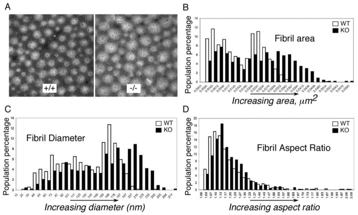
Figure 2. Collagen fibrils in tendon of Thbs4−/− mice.
(A) Transmission electron micrographs (TEMS) of collagen fibrils in patellar tendon of Thbs4−/− (right) and WT (left) mice. Scale bars = 0.5 μm. Histograms of collagen fibril area (B), fibril diameter (C) and fibril aspect ratio (D) in WT mice (open bars, n = 5) and Thbs4−/− mice (black bars, n =5). Fibril areas and fibril diameters are more variable, and larger fibrils are more prevalent in patellar tendon of Thbs4−/− mice but the shapes of fibrils, assessed from the aspect ratio, are not altered.

